The Research Club was instituted by the Laboratory of Vascular Physiology and Medicine under the auspices of the Department of Physiology in the year 2017(January).
Aim: Aimed to promote research culture and research environment in the BLDE University.
Objectives:
The Research Club has been formed with the following objectives
Date, Time & Venue of The Research Club meet:The last Saturday of every month from 4.00 pm to 5.30 pm in the Laboratory of Vascular Physiology and Medicine, Department of Physiology.
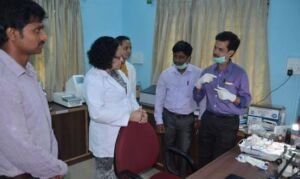
Inception and Inauguration: The 1st meet and formal inauguration of The Research Club was held on 28.01.2017. Prof B. G. Mulimani, Chief Advisor and Former Vice-Chancellor, BLDE (Deemed to be University) inaugurated “The Research Club” and Dr. M. S. Biradar, Former Vice-Chancellor, BLDE (Deemed to be University) presided over the function. Prof Kusal K. Das, Mentor for The Research Club briefed about the objectives of The Research Club and also shared some interesting facts and information about research clubs in other universities of the world. Members of The Research Club shared their views on the club and also their research experience. Since its inception total of 22 Research club meets have been organized (10 in the year 2017 and 12 in the year 2018). Resource Persons specialized in various fields were invited for The Research Club meets. They shared their expertise and views on various topics with the members of the Research Club.
| S. No. | Date | Hot Topic | Popular Topic |
|---|---|---|---|
| 1 | 28th Jan 2017 | Formal inauguration by Prof B. G. Mulimani, Chief Advisor and Former Vice Chancellor, BLDE University and Dr M. S. Biradar, Hon’ble Vice-Chancellor, BLDE University | |
| 2 | 25th Feb 2017 | MoU between Tulane University, New Orleans, US and BLDE University. Prof Kusal K. Das briefed about his experience during visit to Hypertension and Renal Centre of Excellence, Tulane University, New Orleans, US | |
| 3 | 25th Mar 2017 | Hypertension | Science and Philosophy of Music |
| 4 | 22nd Apr 2017 | Cardiomyopathy | Reiki, Summer diet |
| 5 | 27th May 2017 | Organ Transplantation | Laughter therapy |
| 6 | 24th June 2017 | Pathophysiology of Preeclampsia | Gut microbiota |
| 7 | 29th July 2017 | Urinary tract infection | Mediterranean diet |
| 8 | 26th Aug 2017 | Recent advances in diabetes management | Surrogacy: A Legal Perspective |
| 9 | 28th Oct 2017 | Motor neuron disease | Progeria-Old before time |
| 10 | 16th Dec 2017 | Prospects and Future of Physiology | |
| 11 | 23rd Jan 2018 | Science and Religion | |
| 12 | 24th Feb 2018 | Carcinoma of Breast | Smart Phone Hazards |
| 13 | 31st Mar 2018 | Immunotherapy | The theory of everything in memory of Late Prof. Stephen Hawking |
| 14 | 28th Apr 2018 | Oxidative stress and antioxidant defence | Sleep Quality |
| 15 | 26th May 2018 | Significance of citations | Birds in and around Bijapur |
| 16 | 30th Jun 2018 | Biomedical waste management | mHealth |
| 17 | 03 Aug 2018 | Role of human genetics in medical diagnosis | Physical activity and health |
| 18 | 25th Aug 2018 | Importance of medical education on growth of medical university | Health impact of extreme climatic conditions |
| 19 & 20 | 27th Oct 2018 | Dyslexia | Body donation |
| 21 | 24th Nov 2018 | Carcinoma of cervix | Artificial Intelligence in Health Care |
| 22 | 29th Dec 2018 | Cancer counseling | E-cigarettes-Pros and Cons |
| 23 | Quality of drinking water in and around Vijayapur | Are Universities becoming emotionless? | |
| 24 | Importance of pets in human life | Cooking is both physical and mental therapy. | |
| 25 | Human ergonomics in changing scenario for better Indian health care system | Pre INCA civilization in Bolivia. | |
| 26 | How to keep scientific innovation alive | Keto diet | |
| 27 | Neonatal care and infant mortality in and around Vijayapur | Stippling art. | |
| 28 | Research and development in the perspective of peer assessments | ||
| 29 | Interdisciplinary research between Pharmacies especially drug development and Clinical trials with Medicine. | Water crisis and future of India | |
| 30 | Interdisciplinary research with Engineering & Medical Sciences under BLDE umbrella | Human touch at the face of nature’s fury. | |
| 31 | Significance of interdisciplinary research between Western and Indian systems of Medicine. | North Karnataka delicacies. | |
| 32 | Importance of school education in the perspective of higher education. | Global hunger index and India. | |
| 33 | Medical Education and Research in Bolivia. | ||
| 34 | Global warming, climate change, and environmental health. | Happy New Year 2020 | |
| 35 | Environmental Engineering: Scope & Future Area of Research | ||
| 36 | ‘National Science Day 2020 Celebration’ on 28th February 2020 | ||
| 37 | Introduction to Bioinformatics | ||
| 38 | Prof. A. S. Paintal, FRS 94th BIRTH ANNIVERSARY’: 24th September 2020 | ||
| 39 | Molecular design and synthesis | ||
| 40 | Journey of Physiology Education: My Perception | ||
| 41 | Unlocking Physiology, Crossing Borders | ||
| 42 | New perspective of the control of CO2/H+-dependent drive to breathe | ||
| 43 | Impact of erythropoietin on neuronal control of respiration | ||
| 44 | DNA repair System In Mycobacteria | ||
| 45 | Development of Electrochemical Sensing Interfaces by using Advanced Voltammetric Techniques (KOREATECH) |
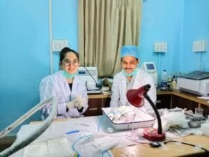
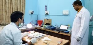
Research Models developed at Vascular Lab
External Research Student/Faculty Training
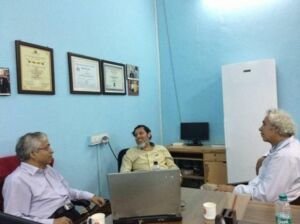
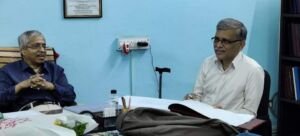
Dr.Naira Eloyan, UNESCO PhD student under UNESCO Chair Life Sciences from Yerevan, Armenia had undergone 3 week training at Vascular Laboratory. She was mentored by Prof.Kusal Das, UNESCO Adviser for Environmental Health and PhD Co-Adviser of Dr.Naira Eloyan. Dr.Vidya A.Patil, Professor & Head, Department of Anaesthesia acted as a clinical mentor for Dr.Naira for clinical research training (September – October 2020).
Dr.Naira was trained in experimental physiology from the perspective of biophysics and molecular biology. She also received clinical training in the Department of Anaesthesia for the management of pain sensation and electromagnetic field.
Dr.Natalia Zubieta-DeUrioste,MD – A physician at La Paz, Bolivia at High Altitude Pulmonary and Pathology Institute visited Laboratory of Vascular Physiology and Medicine, BLDE (Deemed to be University) as a research trainee for 2 weeks during November – December 2020. She had a training basically on experimental physiology and molecular biology specific to hypoxia gene here.
Dr.Satish Dipankar, MD (Physiology), Associate Professor of Physiology, AIIMS, Patna was deputed to the Laboratory of Vascular Physiology and Medicine for research training in experimental physiology under Prof.Kusal Das. He was here for 2 weeks in June 2020. During his training, he learned the rodent stroke model and experimental hypertension.
RESEARCH IMPACT FROM VASCULAR LABORATORY
Collaborative research with University of Leeds(2016) able to successfully established experimental rat cerebral ischemicmodelat Vascular Physiology laboratory of the University. Using this model as preclinical studies vascular science researchers of BLDE (DU) demonstrated clearly that hypoxia pre-treatment (5% O2)in animals can reduce ischemic brain injury due to stroke ! This unique research indicated as a possible protective strategy against cerebral focal ischemia or stroke (Das et al 2018).
Collaborative research on hypoxia and high-altitude pathophysiology with High Altitude Pulmonary and Pathology Institute, Bolivia and Laval University, Canada the researcher of BLDE (DU) demonstrated that the oxygen transport triad (pneumodynamic pump, hemodynamic pump & Epo/Hemoglobin) actually regulates hypoxia tolerance of an individual in relation to physiological acid-base balance. These hypotheses partly explain why COVID-19 incidence is lower in high altitude inhabitants. Further, the work also suggested hypoxia preconditioning as a preventive measure against acute hypoxic complications in high altitude like HAPE or HACE, etc.(Zubieta et al 2020).
1. Zubieta-Calleja, G.R.; Zubieta-DeUrioste, N.; Venkatesh, T.; Das KK.; Soliz, J. COVID-19: Multiple Diseases Simulating Extreme High-Altitude Exposure? Oxygen Transport Physiology and Scarce Need of Ventilators; Andean Condor & rsquo;s-Eye-View. Reviews on Recent Clinical Trials 2020, 15(4).
https://doi.org/10.2174/1574887115666200925141108
Normalized wall index,as an indicator of vascular structural integrity might be useful to assess the atherosclerotic disease burden. It is routinely used in Carotid Doppler study as well as in CT and MRI to measure the wall thickness and lumen diameter of blood vessel in humans. Vascular researchers of BLDE (DU) have made successfully thefirst time an attempt to evaluate the normalized wall index (NWI)through histopathology in experimental rat model by using Digimizer Image analyser (Patil et al 2019).
This innovative technique to measure the arterial wall and lumen thickness through histopathology was first ever reported in any scientific literatureso far by anyone other than BLDE(DU) researchers. This invention will definitely change remarkably in vascular science research for preclinical trials.
Chronic intermittent hypoxia (CIH) is commonly seen in obstructive sleep apnoea. It is associated with cardiovascular diseases, hypertension and impaired glucose homeostasis. Researchers of vascular laboratory of BLDE(DU) showed clearly in experimental model that CIH induces over production of reactive oxygen species as well as hyper activities of sympathetic N-typeCa2+ channels possibly through HIF 1-α expression and influence on insulin signalling by causing hyperglycemia, glucose intolerance and insulin resistance. This hyperglycaemic status can be controlled by using L/N type calcium channel blocker like cilnidipine as possible therapeutic measure(Bagali et al 2020; Das et al 2016).
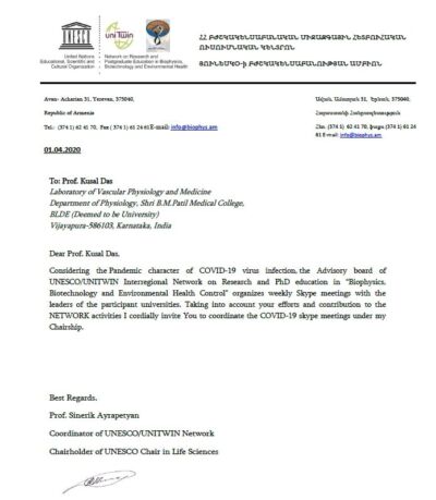
(http://www.biophys.am/pages/index/Environmental-Health/ )
LSRB, DRDO
Ministry of Defence, Government of India (2014- 2017 extended 2019 September ).Title: “Influence of antioxidant vitamin (L-ascorbic acid) on hypoxia-induced alteration of VEGF gene expression in male diabetic rats with or without exposure to heavy metal nickel”. Fund Rs. 27,40,600/-(Vide Sanction letter: No O/o CC R&D(TM)/81/48222/LSRB-XXIX-Meeting/2014 06th August 2014) [Completed]
INR 17, 65,200 (2015-2019)
VGST-K FIST (Level 2),
(DST, Government of Karnataka (2016-2019). “Effect of L-ascorbic acid and calcium channel blocker on hypoxia exposed possible alteration of cell signaling pathways in the respiratory system of male rats with or without heavy metal lead exposure” (VGST-KFIST/1230/2015 Dated 22/6/2016.(Rs.40,00,000/-) [On Going]
INR 40, 00,000 ((2017-2021)
KSTA-GRANT,
for Medical Ethics Seminar (DST. Government of Karnataka, Feb. 2021).
INR 1,00,000. ( 2021)
MP45 BIOPAC STUDENT LAB:
SMALL ANIMAL RADIO TELEMETRY SYSTEM:
UV VISIBLE SPECTROPHOTOMETER (SCHIMADZU UV 1800):
PERISCOPE:
ELISA MICRO-PLATE READER:
ELISA MICRO-PLATE WASHER
WESTERN BLOTTING APPARATUS
PCR ( THERMOCYCLER) MACHINE
To identify a specific gene For gene mutation studies by using restriction fragment length polymorphism (RFLP) technique
COLD CENTRIFUGE (-200 C with 20000 rpm, REMI )
VERTICAL DEEP FREEZER (ELANPRO -20 DEGREE CELSIUS)
DRUG SCREENING (ADME)
MOLECULAR DOCKING WITH ALL THE SOFTWARES
PHYSIO-PAC:
DICROWIN (PC BASED PPG ANALYZER)
CANWIN (CARDIAC AUTONOMIC NEUROPATHY ANALYZER)